


Details
| Product Name | Recombinant Rabbit Anti-NDUFB9 Monoclonal Antibody |
| Cat No. | SLA2500M |
| Chinese Name | NDUFB9蛋白重组兔单抗 |
| Alias | B22; CI-B22; LYRM3; MC1DN24; UQOR22; 1190008J14Rik; NDUB9_HUMAN; NDUFB9; Complex I-B22 (CI-B22); LYR motif-containing protein 3; NADH-ubiquinone oxidoreductase B22 subunit; NDUB9_MOUSE; NADH:ubiquinone oxidoreductase subunit B9; NADH dehydrogenase (ubiquinone) 1 beta subcomplex, 9 (22kD, B22); NADH dehydrogenase (ubiquinone) 1 beta subcomplex, 9, 22kDa; complex I B22 subunit |
| Host Species | Rabbit |
| Isotype | IgG |
| Clonality | Monoclonal |
| Clone No. | 8A6 |
| React Species | Human,Mouse,Rat |
| Applications | WB (1:500-2000) IHC-P (1:100-500) IHC-F (1:100-500) IF (1:100-500) Flow-Cyt (1:50-100) ICC/IF (1:50-200) IP (1:20-50) Not yet tested in other applications. Optimal working dilutions must be determined by the end user. |
| Immunogen | A synthesized peptide derived from human NDUFB9 |
| Purification | affinity purified by Protein A |
| Predicted MW. | 22 kDa |
| Observed MW. | 23 kDa |
| Subcellular Location | N/A |
| Gene ID | 4715 |
| Swiss-Prot | Q9Y6M9 |
| Storage | 0.01M TBS (pH7.4) with 1% BSA, 0.02% Proclin300 and 50% Glycerol. Shipped at 4℃. Store at -20℃ for one year. Avoid repeated freeze/thaw cycles. |
| Background | Accessory subunit of the mitochondrial membrane respiratory chain NADH dehydrogenase (Complex I), that is believed to be not involved in catalysis. Complex I functions in the transfer of electrons from NADH to the respiratory chain. The immediate electron acceptor for the enzyme is believed to be ubiquinone. |
| Attention | This product as supplied is intended for research use only, not for use in human, therapeutic or diagnostic applications. |
Validation Images
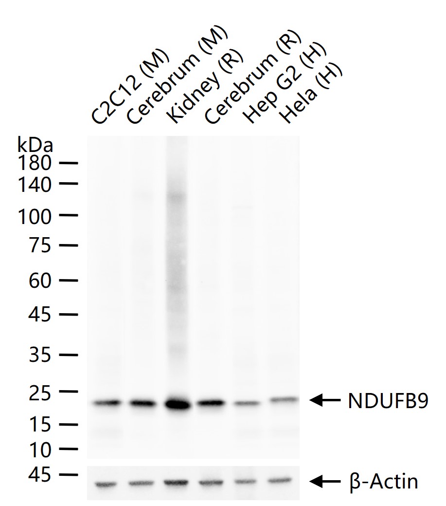
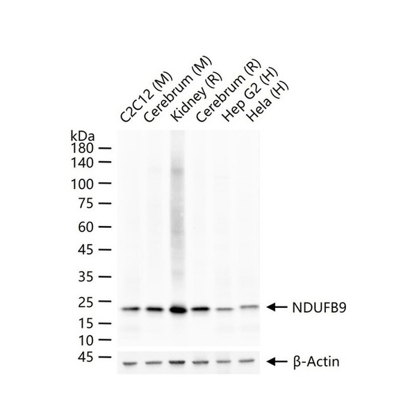
Figure 1. 25 ug total protein per lane of various lysates (see on figure) probed with NDUFB9 monoclonal antibody, unconjugated (SLA2500M) at 1:1000 dilution and 4°C overnight incubation. Followed by conjugated secondary antibody incubation at r.t. for 60 min |
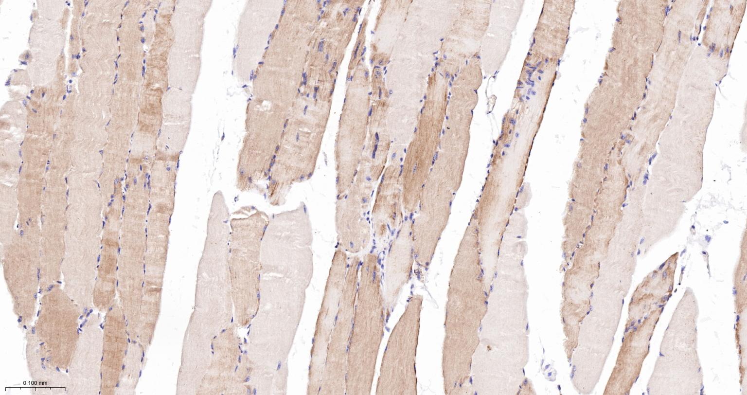
Figure 2. Paraformaldehyde-fixed, paraffin embedded Mouse Skeletal muscle; Antigen retrieval by boiling in sodium citrate buffer (pH6.0) for 15 min; The section was incubated with NDUFB9 Monoclonal Antibody, Unconjugated (SLA2500M) at 1:200 overnight at 4°C, followed by conjugation to the SL0295G-HRP and DAB (C-0010) staining |
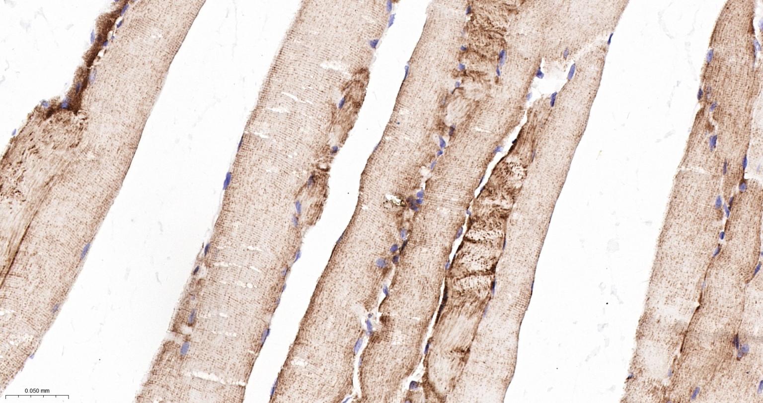
Figure 3. Paraformaldehyde-fixed, paraffin embedded Rat Skeletal muscle; Antigen retrieval by boiling in sodium citrate buffer (pH6.0) for 15 min; The section was incubated with NDUFB9 Monoclonal Antibody, Unconjugated (SLA2500M) at 1:200 overnight at 4°C, followed by conjugation to the SL0295G-HRP and DAB (C-0010) staining |
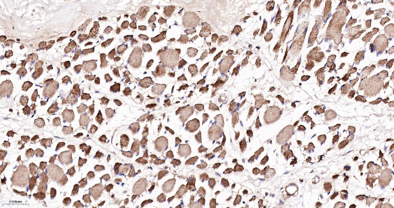
Figure 4. Paraformaldehyde-fixed, paraffin embedded Human Skeletal muscle; Antigen retrieval by boiling in sodium citrate buffer (pH6.0) for 15 min; The section was incubated with NDUFB9 Monoclonal Antibody, Unconjugated (SLA2500M) at 1:200 overnight at 4°C, followed by conjugation to the SL0295G-HRP and DAB (C-0010) staining |
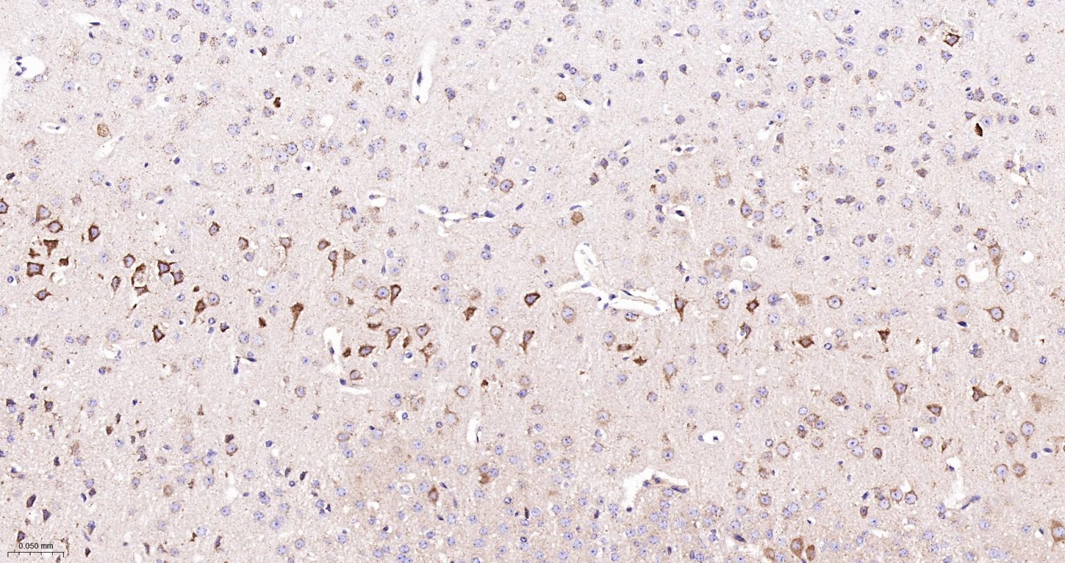
Figure 5. Paraformaldehyde-fixed, paraffin embedded Mouse Cerebrum; Antigen retrieval by boiling in sodium citrate buffer (pH6.0) for 15 min; The section was incubated with NDUFB9 Monoclonal Antibody, Unconjugated (SLA2500M) at 1:200 overnight at 4°C, followed by conjugation to the SL0295G-HRP and DAB (C-0010) staining |
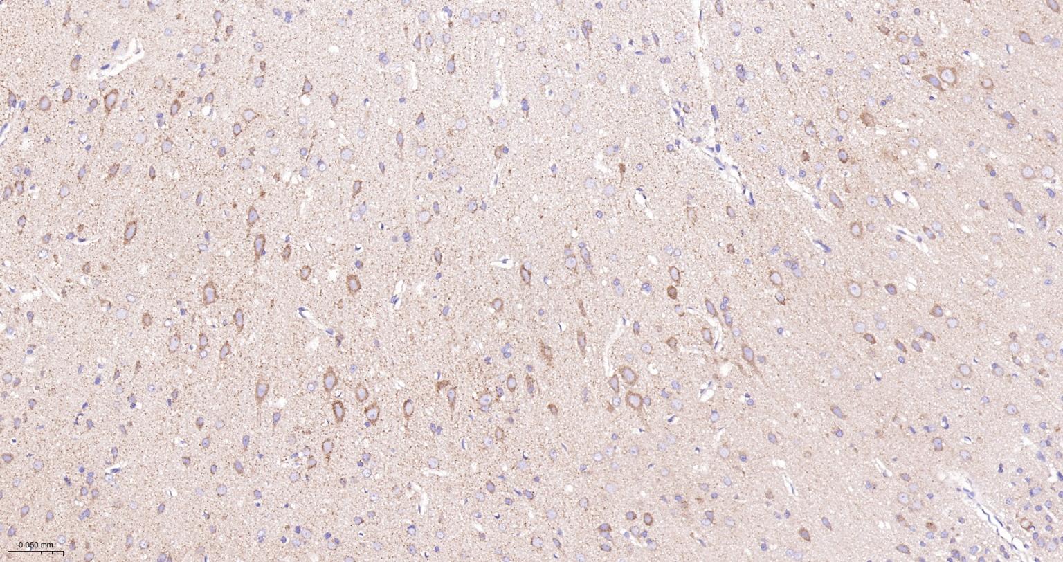
Figure 6. Paraformaldehyde-fixed, paraffin embedded Rat Cerebrum; Antigen retrieval by boiling in sodium citrate buffer (pH6.0) for 15 min; The section was incubated with NDUFB9 Monoclonal Antibody, Unconjugated (SLA2500M) at 1:200 overnight at 4°C, followed by conjugation to the SL0295G-HRP and DAB (C-0010) staining |
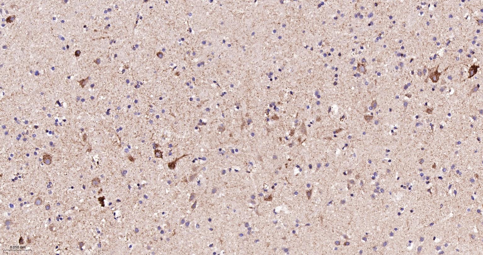
Figure 7. Paraformaldehyde-fixed, paraffin embedded Human Cerebrum; Antigen retrieval by boiling in sodium citrate buffer (pH6.0) for 15 min; The section was incubated with NDUFB9 Monoclonal Antibody, Unconjugated (SLA2500M) at 1:200 overnight at 4°C, followed by conjugation to the SL0295G-HRP and DAB (C-0010) staining |
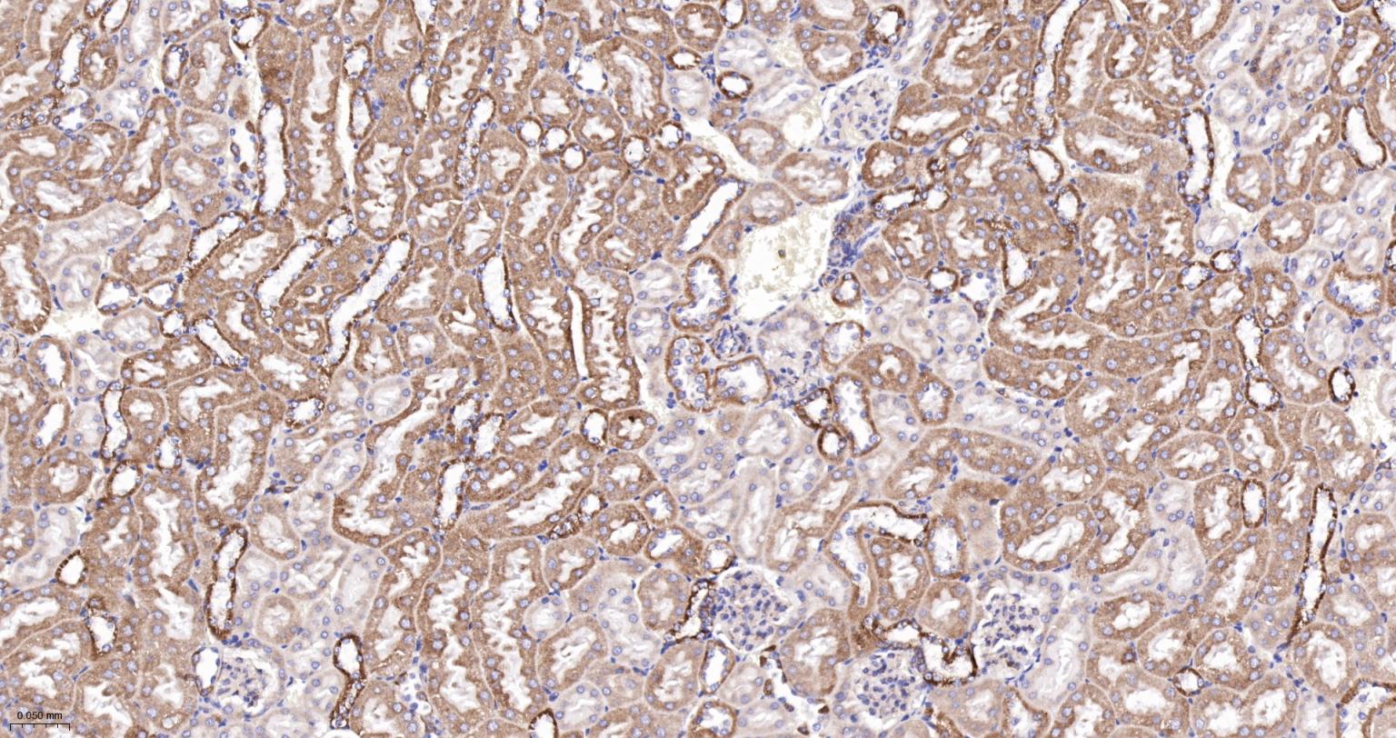
Figure 8. Paraformaldehyde-fixed, paraffin embedded Mouse Kidney; Antigen retrieval by boiling in sodium citrate buffer (pH6.0) for 15 min; The section was incubated with NDUFB9 Monoclonal Antibody, Unconjugated (SLA2500M) at 1:200 overnight at 4°C, followed by conjugation to the SL0295G-HRP and DAB (C-0010) staining |
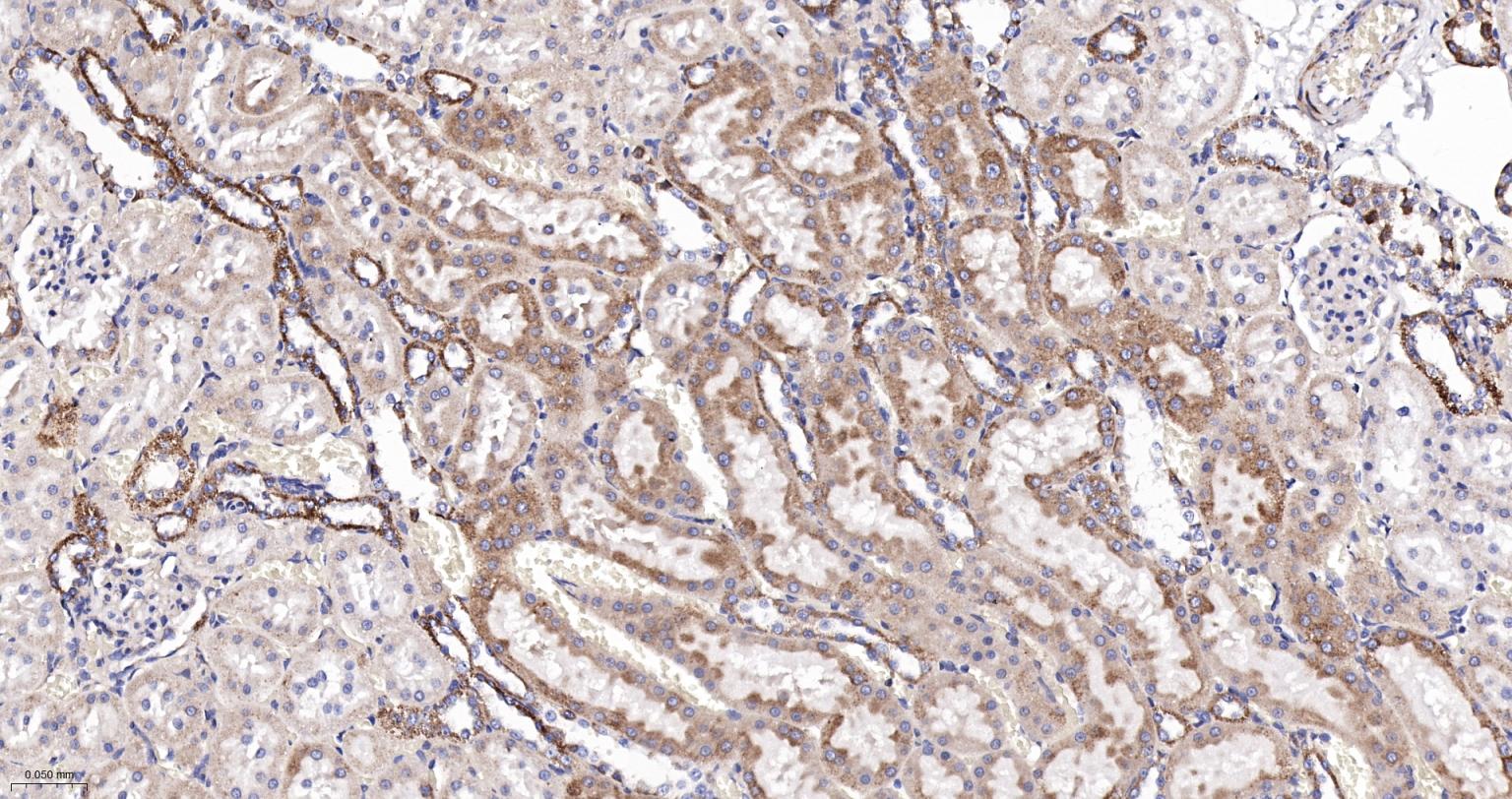
Figure 9. Paraformaldehyde-fixed, paraffin embedded Rat Kidney; Antigen retrieval by boiling in sodium citrate buffer (pH6.0) for 15 min; The section was incubated with NDUFB9 Monoclonal Antibody, Unconjugated (SLA2500M) at 1:200 overnight at 4°C, followed by conjugation to the SL0295G-HRP and DAB (C-0010) staining |
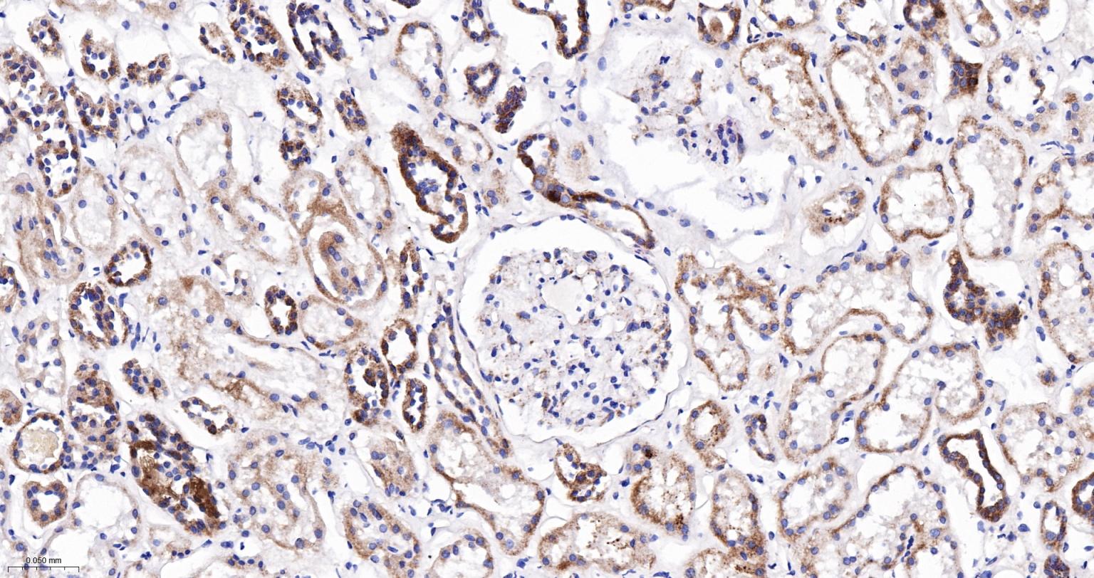
Figure 10. Paraformaldehyde-fixed, paraffin embedded Human Kidney; Antigen retrieval by boiling in sodium citrate buffer (pH6.0) for 15 min; The section was incubated with NDUFB9 Monoclonal Antibody, Unconjugated (SLA2500M) at 1:200 overnight at 4°C, followed by conjugation to the SL0295G-HRP and DAB (C-0010) staining |
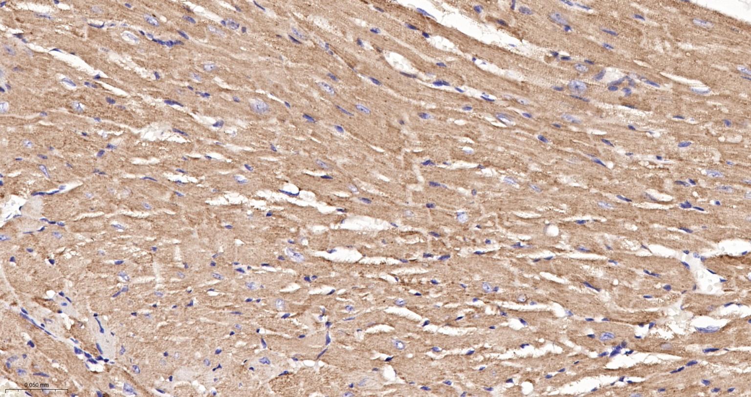
Figure 11. Paraformaldehyde-fixed, paraffin embedded Mouse Heart; Antigen retrieval by boiling in sodium citrate buffer (pH6.0) for 15 min; The section was incubated with NDUFB9 Monoclonal Antibody, Unconjugated (SLA2500M) at 1:200 overnight at 4°C, followed by conjugation to the SL0295G-HRP and DAB (C-0010) staining |
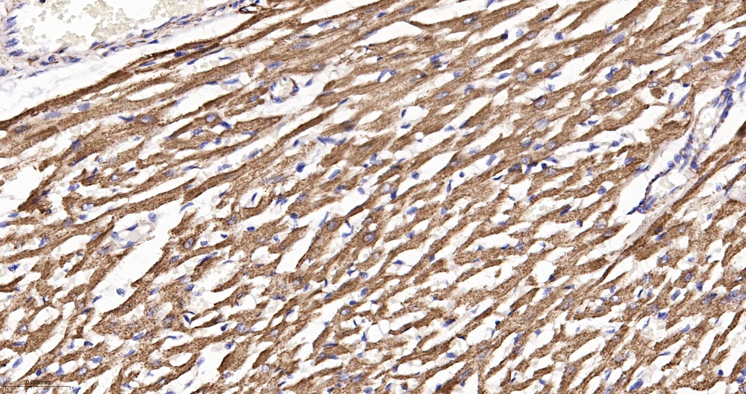
Figure 12. Paraformaldehyde-fixed, paraffin embedded Rat Heart; Antigen retrieval by boiling in sodium citrate buffer (pH6.0) for 15 min; The section was incubated with NDUFB9 Monoclonal Antibody, Unconjugated (SLA2500M) at 1:200 overnight at 4°C, followed by conjugation to the SL0295G-HRP and DAB (C-0010) staining |
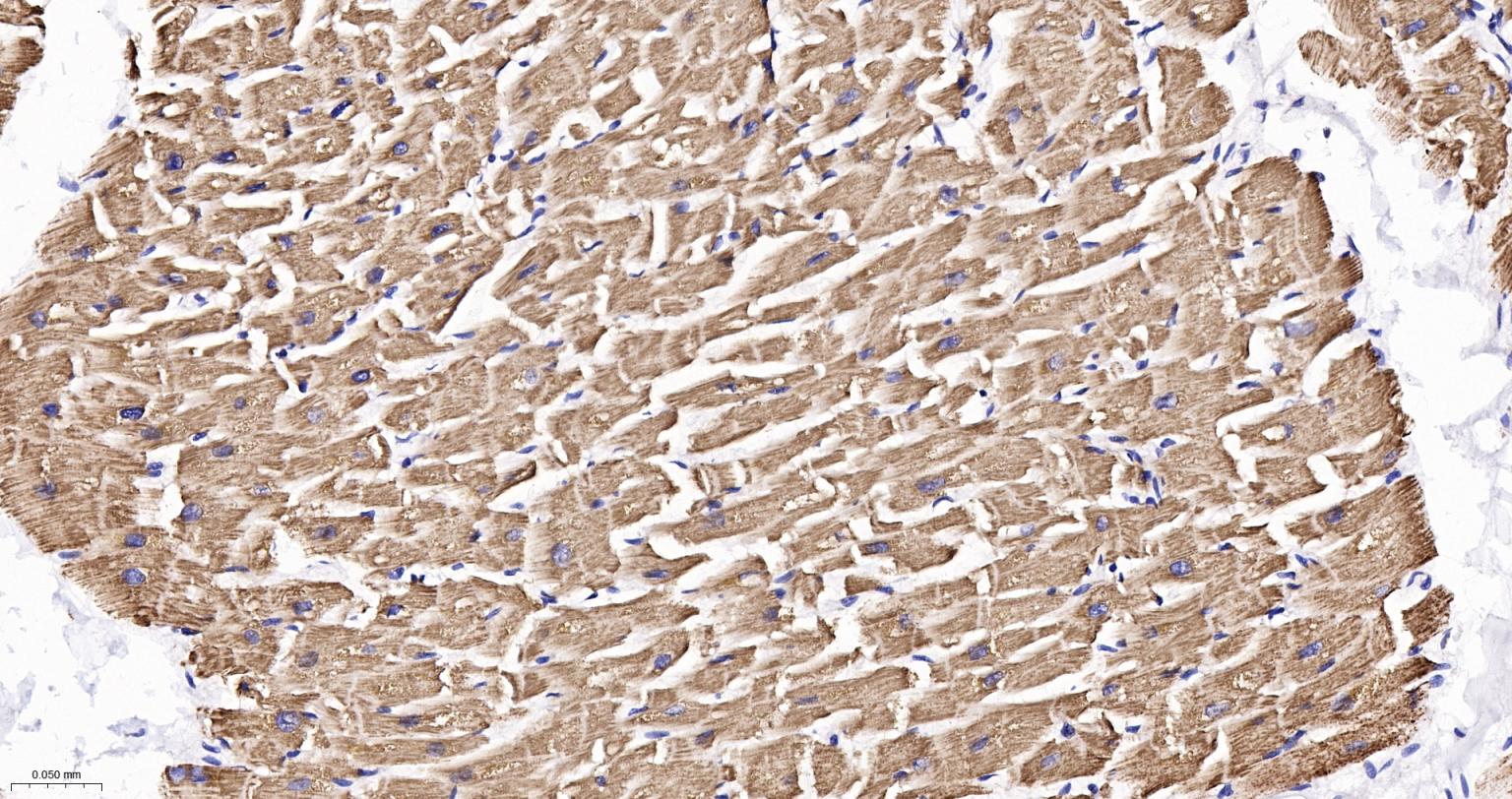
Figure 13. Paraformaldehyde-fixed, paraffin embedded Human Heart; Antigen retrieval by boiling in sodium citrate buffer (pH6.0) for 15 min; The section was incubated with NDUFB9 Monoclonal Antibody, Unconjugated (SLA2500M) at 1:200 overnight at 4°C, followed by conjugation to the SL0295G-HRP and DAB (C-0010) staining |
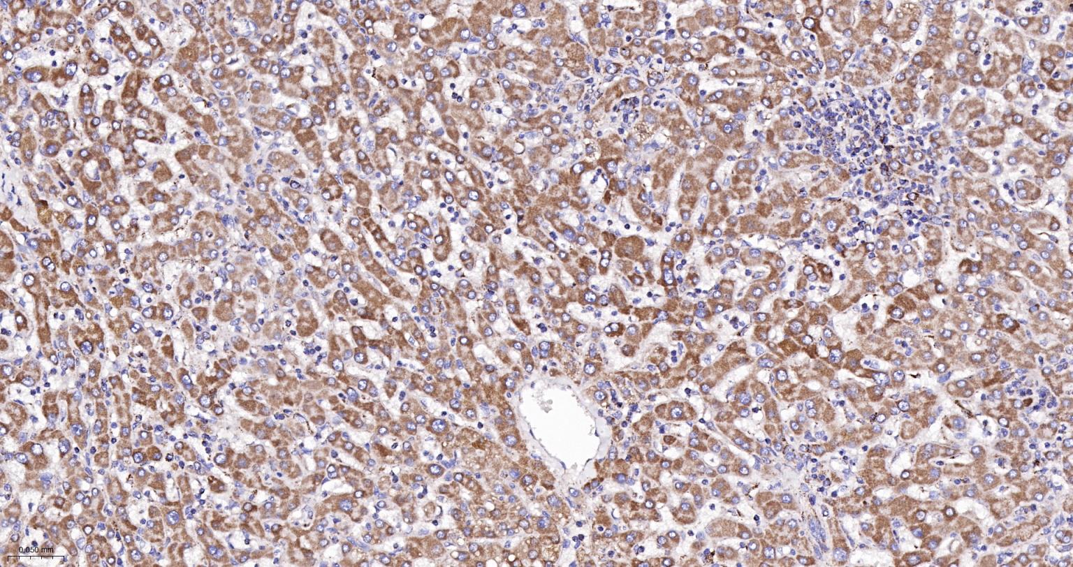
Figure 14. Paraformaldehyde-fixed, paraffin embedded Human Liver; Antigen retrieval by boiling in sodium citrate buffer (pH6.0) for 15 min; The section was incubated with NDUFB9 Monoclonal Antibody, Unconjugated (SLA2500M) at 1:200 overnight at 4°C, followed by conjugation to the SL0295G-HRP and DAB (C-0010) staining |
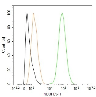
Figure 15. The HepG2 (H) cells were fixed with 4% PFA (10 min at r.t.) and then permeabilized with 90% ice-cold methanol for 20 min at -20℃,the cells then were incubated in 5%BSA to block non-specific protein-protein interactions (30 min at r.t.), followed by secondary antibody incubation for 40 min at room temperature. Primary Antibody (green):Rabbit Anti-NDUFB9 antibody (SLA2500M,1:100); Isotype Control (orange): Rabbit IgG (SL0295P). Blank control (black): PBS. Acquisition of 20,000 events was performed |
FOR RESEARCH USE ONLY. Not for use in diagnostic or therapeutic procedures.
Ordering & shipping
Partial purchase records (0)
Leave a message
Call us
+86 0571 56623320
Address
Room 1-315, Kongle Changqing Building, No. 160 Guangye Road,Gongshu District, Hangzhou City, Zhejiang Province, China
About Us Contact Us Privacy Policy Shipping Policy Payment Policy Quality Guarantee Return & Refund policy Download Product list Distributors


 +86 0571 56623320
 [email protected]
 +86 18668110335


Scan Wechat Qrcode





 Manual
