


Details
Sunlong Medical™ High Five (H5) Host Cell Protein (HCP) G3 Residual Detection ELISA Kit (One-Step)
For research use only. Not for use in diagnostic procedures.
1. APPLICATION
For the quantitative determination of host cell protein (HCP) residues of High Five (H5) cells in protein purification processes and final products. The kit is suitable for in-process and final-product samples and is intended for the monitoring of host cell protein clearance during downstream process development and quality control.
This kit is intended for the quantitative analysis of residual host cell protein (HCP) in protein drugs when High Five (H5) cells, derived from Trichoplusia ni, are used as the expression system.
2. PRINCIPLE
This kit is based on the solid-phase sandwich enzyme-linked immunosorbent assay (ELISA). A capture antibody is pre-coated onto the microplate to form the solid-phase antibody. During the assay, the antigen to be tested and an enzyme-labeled detection antibody are added simultaneously to the coated wells to form a capture antibody-antigen-detection antibody complex. After washing, unbound conjugates are removed, and the substrate is added for color development. Finally, the Stop Solution is added to stop the reaction. The color intensity is positively correlated with the total protein content. The absorbance (OD value) is measured on a microplate reader at 405 nm, fitted against the concentration to construct a standard curve, and the sample HCP concentration is calculated by substituting the sample OD value into the standard curve equation.
3. KIT SPECIFICATIONS
Sensitivity (LOD): 3.125 ng/mL
Limit of quantification (LOQ): 12.5 ng/mL
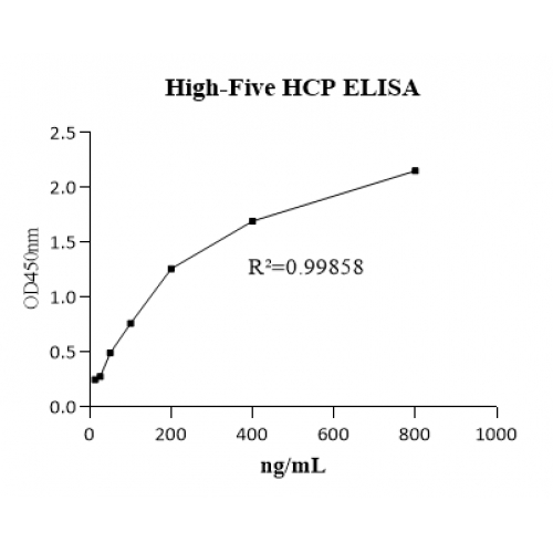
Assay range: 12.5 - 800 ng/mL (7-point standard curve, S1 = 800 ng/mL)
Precision: intra-assay CV < 10%; inter-assay CV < 10%
Sample volume: 50 μL per well
Assay type: solid-phase sandwich ELISA (one-step), read at 405 nm
Specificity: The cross-reactivity of this assay with the following related host cell proteins was evaluated.
| Sample | Concentration |
| HEK293 HCP | < 2% |
| E. coli HCP | < 1.5% |
| Pichia pastoris HCP | Not Detectable |
| Saccharomyces cerevisiae HCP | Not Detectable |
Recovery: sample-matrix dependent; a spike recovery test is recommended when a different buffer is used for the first time, in order to exclude matrix interference effects; acceptable spike recovery 80 - 120%; in practice, mix the diluted standard S1 (800 ng/mL) with the sample matrix at a 1:1 volume ratio
4. MATERIALS PROVIDED
For items that differ between the two kit sizes, values are given as 48 Test / 96 Test.
| # | Component | Specification |
| 1 | Antibody-coated microplate (detachable) | 48 wells / 96 wells |
| 2 | Standard stock solution (500 μg/mL) | 30 μL / 50 μL |
| 3 | Detection Antibody stock (100×) | 100 μL / 150 μL |
| 4 | Substrate | 10 mL |
| 5 | Stop Solution | 10 mL |
| 6 | Wash Buffer Concentrate (20×) | 10 mL |
| 7 | Diluent (10×) | 10 mL |
| 8 | Plate Cover Seals | - |
| 9 | Instructions | - |
5. ASSAY PROCEDURE SUMMARY
Step 1. Add 50 μL of each standard or sample and 100 μL of 1× Detection Antibody to the appropriate wells; seal the plate and incubate at room temperature with shaking for 2 hours.
Step 2. Wash the plate 5 times with 1× Wash Buffer.
Step 3. Add 100 μL of Substrate Solution and incubate for about 15 minutes at room temperature in the dark.
Step 4. Add 50 μL of Stop Solution and read the OD value at 405 nm.
6. TYPICAL DATA
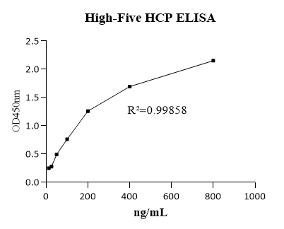
Appendix I: Example of a standard curve (schematic; for reference only).
Ordering & shipping
Partial purchase records (0)
Leave a message
Call us
+86 0571 56623320
Address
Room 1-315, Kongle Changqing Building, No. 160 Guangye Road,Gongshu District, Hangzhou City, Zhejiang Province, China
About Us Contact Us Privacy Policy Shipping Policy Payment Policy Quality Guarantee Return & Refund policy Download Product list Distributors


 +86 0571 56623320
 [email protected]
 +86 18668110335


Scan Wechat Qrcode





 Manual
