


Details
Product Introduction:
Eosinophils are one of the components of inflammatory cells and play a role in allergic diseases. Eosinophils contain secondary granules, which mainly contain basic proteins, such as eosinophil cation, eosinophil derived neurotoxin, eosinophil peroxidase, etc., which can be stained with acid dyes.
Eosinophil staining solution (chromochromic acid 2R method) consisted of hematoxylin staining solution and chromochromic acid 2R staining solution. After staining, the cytoplasm of eosinophils is magenta, and the nucleus is blue. Compared with other eosinophil staining solutions, the color is moderate, the operation is simple and the staining is fast. The disadvantage is that dry slide staining may occur if the tissue patch is not firm.
Note:Product information may be optimized and upgraded. Please refer to the actual label information for accuracy.
Experiment picture
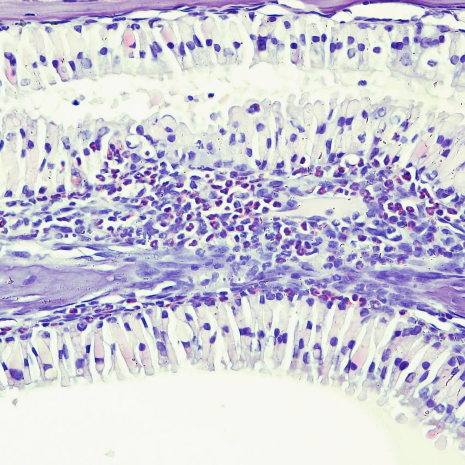
Paraffin section of mouse nasal bone
Eosinophilic granules stain magenta; cell nuclei stain blue; red blood cells stain light yellow or slightly red; eosinophilic tissues such as cartilage stain violet-blue.
Partial purchase records (0)
Leave a message
Call us
+86 0571 56623320
Address
Room 1-315, Kongle Changqing Building, No. 160 Guangye Road,Gongshu District, Hangzhou City, Zhejiang Province, China
About Us Contact Us Privacy Policy Shipping Policy Payment Policy Quality Guarantee Return & Refund policy Download Product list Distributors


 +86 0571 56623320
 [email protected]
 +86 18668110335

Scan Wechat Qrcode





 Manual