


Details
Product Introduction:
The Giemsa stain is composed of azure and eosin. The chemical properties of various cellular components are different, and their affinity for various dyes is also different. Eosinophilic granules are basic proteins that bind to the acid dye eosin and are stained pink; Basophilic granules such as nuclear proteins or lymphocyte cytoplasm are acidic substances, which are stained violet blue by combining with basic dyes methylene blue or azulean; The neutral particles in the isoelectric state can bind to both eosin and methylene blue, showing a light purple color. Each component can be distinguished by combining its own characteristics with different substances in Giemsa staining solution to show different colors.
The Giemsa staining solution prepared by our company is prepared with the imported Giemsa pigment dye as raw material, which can stain the nucleus into purple or blue purple, and the cytoplasm into pink, showing a clear cell staining image under a light microscope. It is mainly used to display the differences in the shape and size of various blood cells in blood smears, with good staining effect, strong staining power and clear staining.
Note:Product information may be optimized and upgraded. Please refer to the actual label information for accuracy.
Experiment picture
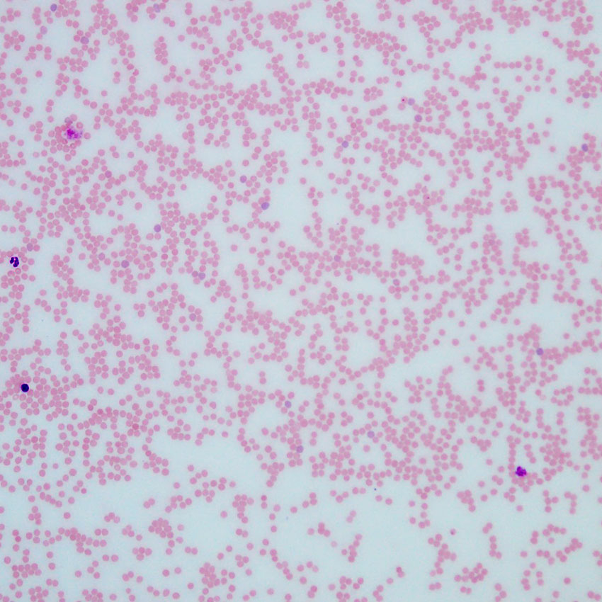
Fresh whole blood smear of mouse
Red blood cells appear light red, slightly paler in the center, giving a disc-like appearance. Lymphocyte membranes stain blue-violet; lymphocyte nuclei show varying shades of purplish-red; lymphocyte cytoplasm stains light red with clear granules.
Partial purchase records (0)
Leave a message
Call us
+86 0571 56623320
Address
Room 1-315, Kongle Changqing Building, No. 160 Guangye Road,Gongshu District, Hangzhou City, Zhejiang Province, China
About Us Contact Us Privacy Policy Shipping Policy Payment Policy Quality Guarantee Return & Refund policy Download Product list Distributors


 +86 0571 56623320
 [email protected]
 +86 18668110335

Scan Wechat Qrcode





 Manual