


Details
Product Introduction:
Collagen fiber is the most widely distributed and abundant fiber in connective tissue. It is widely distributed in various organs. Type I collagen fibers are mainly bone, skin and tendon; Type Ⅱ collagen fibers are mainly cartilage collagen; Type Ⅲ collagen fibers are mainly found in embryonic tissues, adult blood vessels and gastrointestinal tract; Type Ⅳ collagen fibers are mainly in the basement membrane. Sirius red and its lining dye solution are strong acid dyes, which are easy to combine with the basic groups in collagen molecules and have strong adsorption. Polarized light microscopy shows that the collagen fibers have the property of positive uniaxial birefringence. When combining with Sirius red compound staining solution, it could enhance the birefringence and improve the resolution, so as to distinguish the two types of collagen fibers.
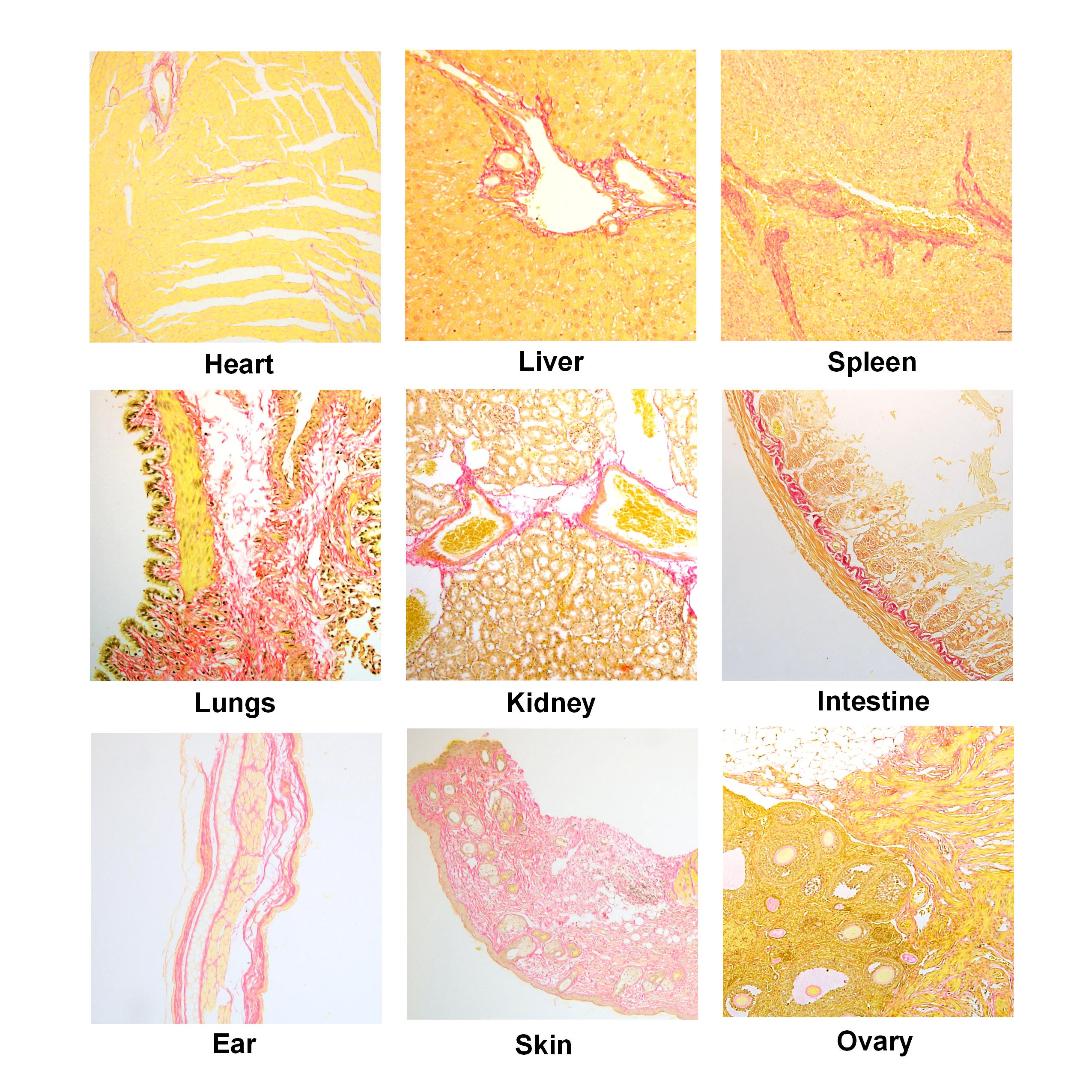
Modified Sirius Red Stain Kit(No Picric Acid) is mainly composed of Iron Hematoxylin Stain and Sirius Red Stain, without picric acid. It is mainly used in the study of collagen fiber abnormality or fiber proliferation in various tissue diseases. Under the ordinary optical microscope, the collagen fibers of heart and blood vessels and other tissues are dyed red, which is helpful for the classification and grading of various fibrotic diseases under the polarized light microscope. Immunohistochemical technique can also display type Ⅰ and Ⅲ collagen fibers, but the antibodies used are expensive and time-consuming, while Sirius red staining reagent is cheap and easy to operate.
Note:Product information may be optimized and upgraded. Please refer to the actual label information for accuracy.
Experiment picture
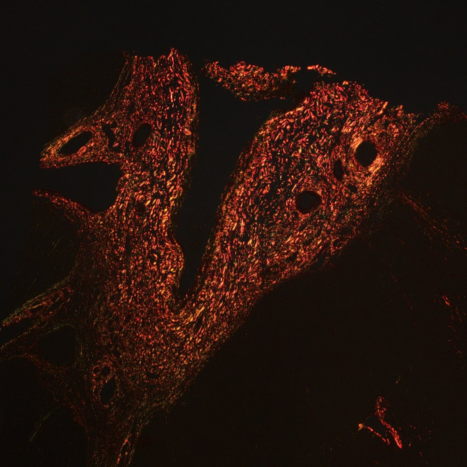
Experiment: Paraffin section of liver cancer – Polarized light microscopy Collagen fibers show birefringence under polarized light: Type I collagen fibers appear strong orange-yellow or bright red; Type III collagen fibers appear green.,
Experiment picture
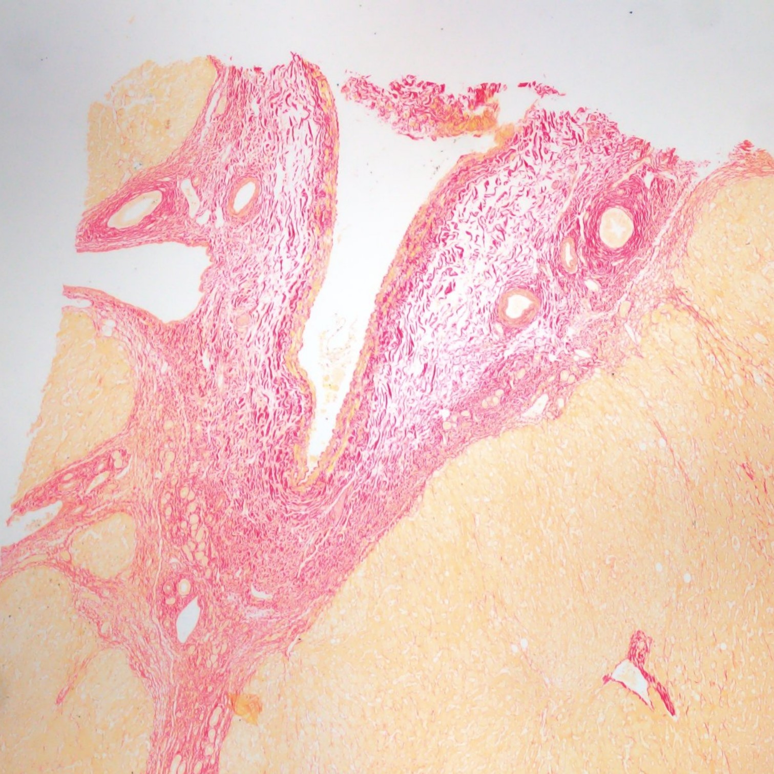
Paraffin section of liver cancer – Light microscopy Collagen fibers stain red; background stains yellow; cell nuclei stain reddish-brown to black.
Partial purchase records (0)
Leave a message
Call us
+86 0571 56623320
Address
Room 1-315, Kongle Changqing Building, No. 160 Guangye Road,Gongshu District, Hangzhou City, Zhejiang Province, China
About Us Contact Us Privacy Policy Shipping Policy Payment Policy Quality Guarantee Return & Refund policy Download Product list Distributors


 +86 0571 56623320
 [email protected]
 +86 18668110335

Scan Wechat Qrcode





 Manual