


Details
| SAMPLE TYPE | Serum, plasma, DMEM cell culture supernatant |
|---|---|
| SAMPLE VOLUME | Serum or plasma: 20μL; DMEM cell culture supernatant: 50μL |
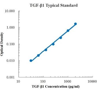
| SENSITIVITY | 9.20 pg/mL |
| RANGE | 15.63 pg/mL - 1000 pg/mL |
| ASSAY TIME | 1.5 h |
| RECOVERY | 88% – 114% |
| AVERAGE RECOVERY | 0.99 |
| PLATFORM | ELISA |
| PLATE | Detachable 96-well plate |
| SIZE | 96T/48T |
| STORAGE | If the reagent kit is unopened, it should be stored at 4℃. However, if it has been opened, the standard solution should be stored at -20℃, while the other components should be stored at 4℃. |
| DELIVERY | 4℃ blue ice transportation |
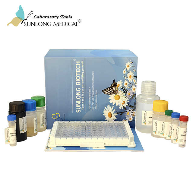
| COMPONENTS | 96-well polystyrene enzyme-linked immunosorbent assay (ELISA) plate coated with anti-TGF-β1 monoclonal antibody TGF-β1 freeze-dried standard TGF-β1 detect Antibody Standard Diluent Assay Buffer(10×) Substrate TMB Stop Solution Washing Buffer(20×) Sealing Film |
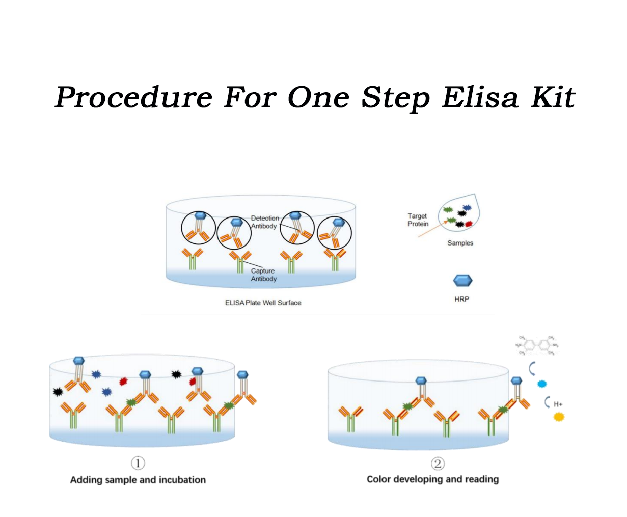
| ASSAY PRINCIPLE | This assay employs the quantitative sandwich enzyme immunoassay technique. Amonoclonal antibody specific for TGF-β1 has been immobilized onto microwells, and one pellet of the HRP-linked detect antibody specific for TGF-β1 (light yellow) is pre-placed in the microwells, sealed by the adhesive film. Standard or samples are pipetted into the wells, then TGF-β1 present is bound by the immobilized antibody and detect antibody. After washing, substrate solution reacts with HRP and color develops in proportion to the amount of TGF-β1 bound by the immobilizedantibody. The color development is stopped and the intensity of the color is measured bymicroplate reader. |
Partial purchase records (2)
| Username | Quantity | bought time |
| Fi*** | 2 | 2024-08-13 |
| Um*** | 3 | 2024-03-11 |
Leave a message
Call us
+86 0571 56623320
Address
Room 1-315, Kongle Changqing Building, No. 160 Guangye Road,Gongshu District, Hangzhou City, Zhejiang Province, China
About Us Contact Us Privacy Policy Shipping Policy Payment Policy Quality Guarantee Return & Refund policy Download Product list Distributors


 +86 0571 56623320
 [email protected]
 +86 18668110335

Scan Wechat Qrcode

