Details
| SAMPLE TYPE | Serum, plasma, DMEM cell culture supernatant |
|---|---|
| SAMPLE VOLUME | Serum or plasma: 10μL; DMEM cell culture supernatant: 33μL |
| SENSITIVITY | 1.77 pg/mL |
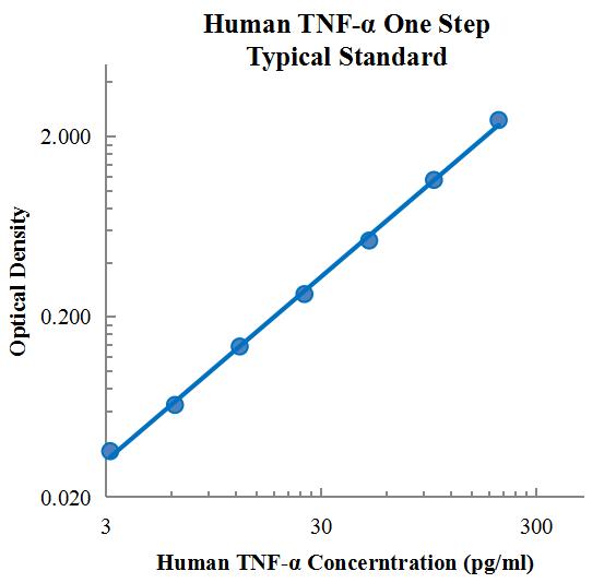
| RANGE | 3.13 pg/mL – 200 pg/mL |
| ASSAY TIME | 1.5 h |
| RECOVERY | 100% – 122% |
| AVERAGE RECOVERY | 1.08 |
| INTRA PRECISION | 4.2% – 5.4% |
| INTER-PRECISION | 5.5% – 6.2% |
| PLATFORM | ELISA |
| PLATE | Detachable 96-well plate |
| SIZE | 96T/48T |
| STORAGE | If the reagent kit is unopened, it should be stored at 4℃. However, if it has been opened, the standard solution should be stored at -20℃, while the other components should be stored at 4℃. |
| DELIVERY | 4℃ blue ice transportation |
| COMPONENTS | 96-well polystyrene enzyme-linked immunosorbent assay (ELISA) plate coated with anti-TNF-α monoclonal antibody Human TNF-α freeze-dried standard TNF-α detect Antibody Standard Diluent Assay Buffer(10×) Substrate TMB Stop Solution Washing Buffer(20×) Sealing Film |
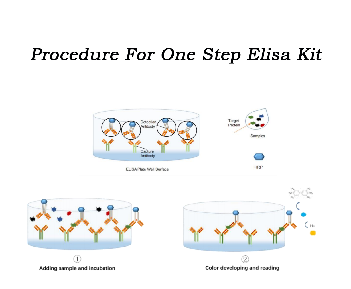
| ASSAY PRINCIPLE | This assay employs the quantitative sandwich enzyme immunoassay technique. Amonoclonal antibody specific for human TNF-α has been immobilized onto microwells, and two pellets of the biotin-linked detect antibody specific for TNF-α (light yellow) and streptavidin-HRP (purple) are pre-placed in the microwells, sealed by the adhesive film. Standard or samples are pipetted intothe wells, then TNF-α present is bound by the immobilized antibody and detect antibody, of whichthe latter is conjugated with streptavidin-HRP in the incubation. After washing, substrate solutionreacts with HRP and color develops in proportion to the amount of TNF-α bound by the immobilized antibody. The color development is stopped and the intensity of the color is measuredby microplate reader. |


Partial purchase records (24)
| Username | Quantity | bought time |
| Is*** | 2 | 2024-09-03 |
| Ma*** | 2 | 2024-08-09 |
| Ta*** | 2 | 2024-08-04 |
| Ca*** | 3 | 2024-08-04 |
| Di*** | 3 | 2024-07-29 |
Leave a message
 +86 0571 56623320
 [email protected]

Scan Wechat Qrcode


Scan Whatsapp Qrcode
